
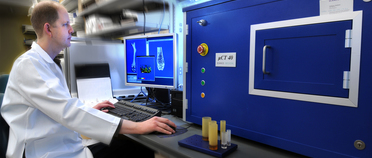
The Scanco40 MicroCT scanner located at Baylor College of Medicine allows for visualization, measurement, and quantification of the structure of bone. This system creates a three-dimensional model from a large number of two-dimensional X-ray images taken around a single axis of rotation.
The high-resolution scanner can capture detail at 6 microns, although a typical scanning resolution of 12 microns is sufficient for most applications. Spines, skulls, and whole mice skeletons can be scanned and used for phenotypic analyses. Upon completion of a scan and reconstruction, the samples can be analyzed or simply viewed to examine morphological features.
The standard types of analysis available are cortical and trabecular analysis, yielding the total volume, bone volume, difference, trabecular thickness, number of trabeculae, trabecular separation, connectivity, and other measurements. This scanner functions as part of a bone analysis core.
Click here for more information about the Scanco40 Instrument at BCM
Specific questions? Please contact Oscar Ruiz (Oscar.Ruiz@bcm.edu)